Olecranon Osteotomy
This is the colour illustration of my self dissected specimen.
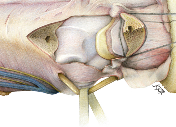
It shows an olecranon osteotomy of a left arm. It was quite difficult to dissect it and to get the perspective right .
This is the colour illustration of my self dissected specimen.
It shows an olecranon osteotomy of a left arm. It was quite difficult to dissect it and to get the perspective right .

First the black and white drawing (aka "sketch") of my self-dissected specimen. We need that to determine were the light is coming from, before we start with the colour illustration. This way you have a reference if you get confused with the warm and cold or bright and dark colours and how they interact with the light.

This is obviously a specimen but in real life you do this when somebody broke something in the joint.
You would think, why saw another bone and 'break' it like the first one?
Why not just approach the broken parts (like the radius or the humerus in the joint) from the other side?
As you can see here, there are nearly no important soft parts like veins, nerves or arteries around the area of the joint. The only important one is easy to secure (it is the N. Ulnaris which is pulled down here). But on the inside of the ellbow joint you have all that. So it is easier for a surgeon to saw trough a bone than operate through a net of very important soft parts.